Abstract
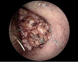
We compare the results of clinical observation and histopathology analysis for developing a differential diagnosis of seborrheic keratosis (SK) of the external auditory canal (EAC). A 46-year-old man with a history of a recurrent lesion in the EAC underwent clinical observation of the skin lesion’s appearance, computed tomography (CT) scan, magnetic resonance imaging (MRI), and several biopsies. Initially, a benign form of SK was diagnosed based on several biopsies performed over a 10-year period. The lesion’s appearance was consistent with a malignant disease, which led the clinician to perform a CT scan and an MRI scan. The patient underwent partial petrosectomy to completely remove the lesion as CT and MRI scans showed an infiltrative process. Squamous carcinoma was the final histological diagnosis. The patient was disease free at 1 year of follow-up after petrosectomy. In conclusion, if there are inconsistencies between clinical observation and histological report, additional tests should be performed to exclude the malignity of a lesion.
Cite this article as: Di Stadio A, Amadori M, Dipietro L, Colangeli R, Falcioni M, Ricci G, et al. Seborrheic Keratosis or Squamous Carcinoma? Clinical Examination versus Biopsy: The Importance of Criticism. J Int Adv Otol 2019; 15(2): 326-9.


.jpg)
.png)
.png)
