
.jpg)
.png)
.png)
Abstract
OBJECTIVES: This study aimed to compare the right and left sides of the carotid foramen (CF) to determine its precise location according to certain anatomical landmarks.
MATERIALS and METHODS: Twenty human dry skulls were included in the study. A digital caliper and a digital image analysis software were used to obtain direct anatomical numerical values. Then, the same parameters on dry skulls were assessed with computed tomography (CT).
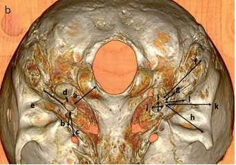
RESULTS: CF was found to be round shaped (62.5%), oval shaped (32.5%), and tear-drop shaped (5%). In all cases, the position of CF was seen as just postero-laterally of the foramen lacerum. According to the jugular foramen, CF was seen to be anterior in 85% and antero-medial in 15% of the cases. Regarding the morphometric values of the surface area, the length and width of CF were observed to be 37.86±11.24 mm2, 8.02±1.09 mm, and 6.86±0.90 mm at direct anatomical measurements and 39.69±10.07 mm2, 7.89±1.14 mm, and 6.41±0.90 mm at CT, respectively. The angles between the supramastoid crest-CF-zygoma root and the supramastoid crest-CF-mastoid process were determined as 37.11±6.87º and 42.22±6.40º at direct anatomical measurements and 36.59±4.94º and 43.71±4.55º at CT, respectively.
CONCLUSION: A significant difference in sides was not observed in relation with the numerical data of CF obtained from CT or from direct anatomical measurements of dry skulls. Moreover, a significant difference was not found between radiological and direct anatomical measurements. Therefore, precise radiological assessment of this region by an experienced neuroradiologist may be assumed as a fundamental need for successful surgeries of the skull base, in addition to thorough anatomical knowledge of neurootologists and neurosurgeons.
Cite this article as: Özalp H, Beger O, Erdoğan O, Koç T, Kayan G, Hamzaoğlu V, et al. Morphometric Assessment of the Carotid Foramen for Lateral Surgical Approach. J Int Adv Otol 2019; 15(2): 222-8.